Search by Blog
Read by Category

Your healing journey continues beyond this blog

Find the right products for better recovery at online store
Cart Image
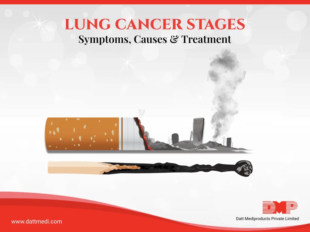
Know the Stages of Lung Cancer

Do you know that despite the advancements in the treatment options, lung cancer remains the leading cause of cancer deaths worldwide for men and the second leading cause for women?

In developed nations, lung cancer is the most common cause of cancer death among women (210,000 deaths) followed by breast cancer (198,000 deaths).

In our last blog, we discussed the 2 types of lung cancer, its complications, risk factors and how to reduce them.

In this blog, we have tried to cover the various stages of Non-small cell lung cancer (NSCLC), which contributes to 85% of the lung cancer types, the various diagnostic methods, and the available treatment options.

Stages of Lung Cancer:

The 2 different types of lung cancer spread in different stages. The different stages help to identify how far the tumor has spread and the treatment to be followed.

Following are the 4 different stages of non-squamous cell lung cancer:

STAGE 1: Cancer is confined to the lungs only and has not yet spread.
STAGE 2: The cancer spreads to the nearby lymph nodes.
STAGE 3: The tumorous cells are found in the lungs, lymph nodes in the middle of the chest. This stage may further be sub-divided into 2 stages based on the side where cancer spreads.
In Stage 3A, The cancer spreads to the lymph nodes which are on the same side of the chest where cancer originated while in Stage 3B, cancer spreads to the opposite side or to the lymph nodes above the collarbone.
STAGE 4: Cancer cells spread to both the lungs or to the surrounding area or to the distant organs.

SCLC Spreads in 2 main stages. In the “limited stage”, cancer is confined to one side of the chest, including one part of the lung and nearby lymph nodes. In the “extensive stage”, cancer cells spread to other parts of the body and the chest.

Diagnosis:
Diagnosis plays an important role, as patient outcomes can improve if the disease is diagnosed at an early stage. However, it is difficult to identify lung cancer at early stages because its symptoms may overlap with those of respiratory infection.

After a physical examination, the doctor may recommend the following diagnostic tests:
– IMAGING TESTS which help to identify any abnormal growth of cells. These may include X-ray, MRI, CT, and PET scans.
– SPUTUM CYTOLOGY, a microscopic examination can help to determine the presence of cancer cells. This may be done if you produce phlegm on coughing.
– BIOPSY may be done to determine if the abnormal growth is cancerous. If the pathological analysis comes positive, further testing such as a bone scan can be done to identify if cancer has spread or not and its stage.

Treatment:
The various treatment options available for lung cancer include surgery, radiation therapy, chemotherapy, radiosurgery, immunotherapy, targeted drug therapy, and palliative care.

SURGICAL METHODS include removing lung cancer and a margin of healthy tissue. Surgery is an option you need for stage 1 NSCLC.

Chemotherapy is often used post surgery to destroy any remaining cancer cells or in patients who are at a high risk of recurrence. Its usually recommended for stage 2 NSCLC and in combination with surgery and radiation therapy in stage 3 NSCLC.

– It is difficult to control stage 4 NSCLC as the cancer cells have spread enough. Treatment may include surgery, radiation, chemotherapy, targeted therapy, and immunotherapy or a combination of these.

– In more advanced stages, where some patients choose not to continue the treatment, palliative care may still help to relieve the cancer symptoms rather than treating cancer itself.

We @DattMediproducts understand that there are no obvious symptoms of the disease at early stages and the diagnosis comes into picture only once cancer has spread.

However, there are ways in which we can reduce the risk. You can follow our previous blog to know more about the complications, risks and how to reduce them.

Visit www.dattmedi.com for more infomation.
Share:
Recent Blogs
Ensuring Safe Wound Care Practices

Wound care is a critical aspect of healthcare that demands meticulous attention to patient safety. Proper wound management not only accelerates healing but also significantly reduces the risk of complications. In this blog, we will explore the importance of patient safety in wound care, emphasizing best practices, infection control, and risk minimization.

Importance of Patient Safety in Wound Care

Patient safety in wound care involves implementing measures to prevent harm and ensure safety during the treatment process. This includes avoiding infections, minimizing pain, and ensuring that wounds heal properly. A safe wound care environment is essential for patient well-being and can prevent further medical complications.

Best Practices in Wound Care

Assessment and Planning:

  • Thorough Assessment: Begin with a comprehensive assessment of the wound, considering factors like wound type, exudate characteristics, comorbid conditions, pain level, periwound condition, size, depth, and location. Understand the patient’s overall health, including any underlying conditions that may affect healing.
  • Individualized Care Plan: Develop a tailored care plan that addresses the specific needs of the patient and the wound. Regularly update the plan based on the wound’s progress.

Hygiene and Infection Control:

  • Clean Environment: Ensure the wound care area is clean and sanitized. All reusable wound care equipment and supplies should be sterilized before use. Fresh dressings and bandages should be cut with clean apparatus.
  • Hand Hygiene: Proper hand hygiene is crucial. Perform hand hygiene using alcohol-based rub or soap and water immediately before and after the wound care procedure. The use of gloves and other personal protective equipment (PPE) is mandatory to prevent contamination.

Wound Cleaning and Dressing:

  • Gentle Cleaning: Clean the wound gently with saline solution or appropriate antiseptic solutions. Avoid harsh scrubbing which can damage tissue and impede healing.
  • Appropriate Dressings: Use the right type of dressing for the wound such as hydrocolloid dressings for pressure ulcers or alginate dressings for heavily exuding wounds. Change dressings as per the doctor’s care plan to maintain a clean environment.

Patient Education and Involvement:

  • Educate Patients: Instruct patients and caregivers on proper wound care techniques, signs of infection, and when to seek medical advice. Patient education enhances compliance and promotes faster healing.

Infection Control:

Preventing infections is a cornerstone of safe wound care. Infections can complicate wound healing and lead to serious health issues. Implementing stringent infection control measures is essential.

  • Sterile Techniques: Always use sterile wound care equipment and supplies when handling wounds. This includes using sterile dressings, tools, and gloves.
  • Regular Monitoring: Monitor the wound for signs of infection, such as increased redness, swelling, warmth, or discharge. Early detection and intervention can prevent severe complications.
  • Antibiotic Use: Use antibiotics judiciously. Overuse or not finishing the full course of the antibiotic can lead to antibiotic resistance, making infections harder to treat.

Minimizing Risks:

Risk minimization in wound care involves anticipating potential problems and taking steps to avoid them.

  • Nutrition and Hydration: Ensure patients have adequate nutrition and hydration. A proper diet supports tissue repair and overall health.
  • Pressure Ulcer Prevention: For bedridden patients, repositioning is crucial to prevent pressure ulcers. Use pressure-relieving devices like specialized mattresses or cushions.
  • Moisture Control: Keep the wound environment moist but not wet. Moisture balance prevents the wound from drying out or becoming too wet, both of which can impede healing.

Ensuring safe wound care practices is vital for promoting patient recovery and preventing complications. By adhering to best practices, maintaining strict infection control, and actively involving patients in their care, healthcare professionals can create a safe and effective wound care environment. As we strive for excellence in wound care, let us remember that patient safety is at the heart of healing.

Adhering to international standards like ISO 9001:2015 and ISO 13485:2016 while manufacturing wound care products can further enhance the quality of wound care practices, ensuring that products meet rigorous safety and quality benchmarks. Let us commit to delivering next-gen healing solutions with a steadfast focus on safety and patient well-being.

Advantages of Using Velfix®࿯ T+Pad for Post Surgical Wounds

In the field of post-surgical wound care, innovation plays a crucial role in ensuring optimal healing outcomes and patient comfort. Introducing Velfix® -T+Pad, a groundbreaking solution that redefines the standards of wound dressings. Meticulously designed and crafted, Velfix® -T+Pad stands at the forefront of advanced wound care technology, offering not only healing but also unparalleled comfort and convenience.

Waterproof and Bacterial Barrier Properties:

Among the essential features of wound dressings, waterproofing and bacterial barrier stand out. Velfix® -T+Pad not only meets these requirements but surpasses them. Its impermeable yet breathable film forms a protective shield against water and bacteria, safeguarding the wound from external threats and fostering a hygienic healing environment.

Benefits of Continuous Visual Monitoring: 

The advantage of transparent film dressings is the ability to visually monitor the wound without removing the dressing. This allows healthcare providers to detect early signs of infection: clinicians can quickly spot signs of infection, excessive drainage, or other complications, facilitating prompt intervention. It also aids in exudate management : ensuring that the wound exudate is being effectively absorbed and does not pose a risk to the healing process.

Conforming to Body Contours:

Comfort is paramount, especially in areas prone to movement or swelling. Velfix® -T+Pad addresses this need adeptly. Its flexible design seamlessly conforms to the body’s contours, providing a snug fit without compromising comfort. Whether it’s a joint, a curve, or a crease, Velfix® -T+Pad stays in place, ensuring uninterrupted healing.

Effective Evaporation of Moisture:

Moisture management is critical for wound healing, and Velfix® -T+Pad excels in this aspect. Thanks to its innovative adhesive pattern, it facilitates efficient moisture evaporation from the wound bed. This not only keeps the area dry but also prevents maceration, creating optimal conditions for healing.

Reducing Pain Upon Removal:

Dressing changes can be painful, but Velfix® -T+Pad aims to alleviate this discomfort. Featuring a non-adherent pad, it minimizes trauma upon removal, reducing pain and discomfort for the patient. With Velfix® -T+Pad, dressing changes become a smoother, more tolerable process, enhancing the overall patient experience.

In the realm of post-surgical wound care, excellence is imperative. With its innovative design and advanced technology, Velfix® -T+Pad sets a new standard for wound dressings, offering a comprehensive solution that prioritizes healing, comfort, and convenience. 

From its waterproof and bacterial barrier properties to its transparent film for easy monitoring, Velfix® -T+Pad epitomizes cutting-edge wound care. Trust in Velfix® -T+Pad to deliver not only healing but also peace of mind, ensuring a smoother, swiffer path to recovery for patients worldwide.

×