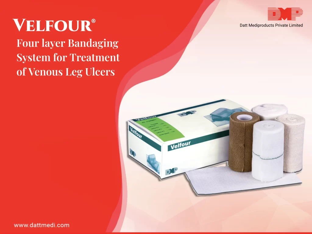
Velfour® is a revolutionary 4-layer compression bandaging system designed to manage venous leg ulcers and associated conditions. With a sub-bandage pressure of 35-40 mm Hg at the ankle, this high-compression system incorporates elastic layers to achieve sustained compression and promote healing.
Velfour® consists of four essential components that work together synergistically to deliver effective compression and support:
1. Velfour®#1 (Orthopaedic wool):
This layer serves as the primary absorbent material, effectively managing exudate while redistributing pressure evenly around the limb.
2. Velfour® #2 (Cotton crepe bandage):
Designed to enhance absorbency, the cotton crepe bandage smooths out the Orthopaedic Wool layer, ensuring optimal comfort and performance.
3. Velfour®#3 (Elastic Compression Bandage):
The first layer of elastic compression sub-bandage, exerting a pressure of approximately 17 mm Hg at the ankle. This layer provides the initial compression necessary for effective treatment.
4. Velfour® #4 (Cohesive Bandage):
The second layer of compression, adding the remaining 23 mm Hg of pressure while ensuring the bandage remains securely in place. The cohesive properties of this bandage contribute to its stability and adherence.
Velfour® offers a range of features and benefits that make it an exceptional choice for venous leg ulcer management:
1. Effective compression:
With a pressure of approximately 40 mm Hg at the ankle, it delivers the necessary compression to promote healing, gradually reducing to 17 mm Hg at the knee. This graduated compression approach ensures optimal therapeutic results.
2. Graduated compression:
The bandage provides high compression at the ankle and gradually decreases the pressure at the calf.
3. Sustained compression:
It maintains a consistent level of compression for up to a full week, allowing for uninterrupted therapy and reducing the frequency of bandage changes. The treatment process is made simpler by this feature, which also increases patient compliance.
The leading option for treating venous leg ulcers, Velfour® also has additional advantages for a number of other ailments, such as:
1. Treatment of Venous Ulcers:
It improves results by hastening the healing process and facilitating the closure of venous leg ulcers.
2. Prevention of Ulcer Recurrence:
In cases where traditional compression stockings are not tolerated, It provides an effective alternative for preventing the recurrence of venous ulcers.
3. Symptomatic Relief of Superficial Thrombophlebitis:
The compression offered by Velfour®alleviates pain and inflammation associated with superficial thrombophlebitis, providing much-needed comfort to patients.
4. Traumatic Wounds with Local Edema:
It aids in reducing edema and promoting the healing of traumatic wounds, helping to restore the affected area to its optimal state.
5. Venous/Lymphatic Disorders:
It is suitable for managing various venous and lymphatic disorders by providing effective compression and support.
To ensure the optimal application and use of Velfour®, it is essential to follow these guidelines:
1. Limb Assessment:
Accurate measurement of the ankle circumference is crucial to determine the appropriate combination of bandages needed. Any changes in limb contour should also be noted for reference.
2. Wound Assessment:
Mapping the ulcer provides a baseline to track progress throughout the treatment. Regular wound assessment enables healthcare professionals to evaluate the effectiveness of Velfour® and make necessary adjustments.
3. Pain Assessment:
This plays a crucial role in managing venous leg ulcers. Compression therapy with it can alleviate pain, although a slight increase may occur initially.
Velfour® stands as a game-changing solution for managing venous leg ulcers. Its innovative 4-layer compression bandage system, combined with its effective pressure distribution and sustained compression, offers unparalleled benefits for patients and healthcare professionals alike.
We are committed to providing valuable information and insights to support your journey towards improved wound healing and patient well-being.
Knee compression supports are one of the most commonly used orthopedic aids — for good reason. They reduce swelling, provide joint stability, and allow people to stay active during recovery. But picking the wrong one delivers less benefit than it should.
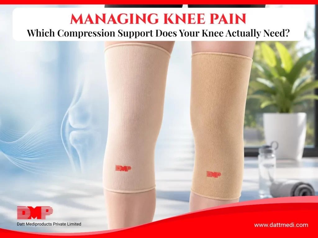
The Datt Mediproducts range includes two knee compression supports: the Knee Cap® and the Knee Cap Classic®. Both share the same anatomically tapered form and four-way elasticized construction, but differ in material composition — and that difference matters for fit, stretch, and the conditions each handles best.
This guide breaks down both products clearly so you can match the right support to the right situation.
Understanding Knee Compression Supports
Before comparing the two products, it helps to understand what knee compression supports actually do — and what they don’t.
A compression sleeve works by applying graduated pressure around the knee joint. This pressure does three things: it reduces fluid accumulation in the surrounding tissue (reducing swelling), it improves preconception — the joint’s sense of its own position in space — which improves stability and coordination, and it provides mild structural support to the muscles and ligaments surrounding the joint.
Compression sleeves are not rigid braces. They do not prevent movement or immobilize the joint. Their role is to support the knee while allowing full, natural range of motion — making them suitable for both active use during daily activity and recovery phases where movement is encouraged.
Knee Cap® vs Knee Cap Classic®: What Is Actually Different?
Both products share the same core design: anatomically tapered, four-way elasticized fabric, breathable moisture-transporting weave, beige color, and easy pull-on application. Available in S to XXL (37–67 cm thigh circumference). The key difference is material composition.
The inclusion of Spandex and Nylon gives the Knee Cap® a higher degree of stretch and elastic recovery. The fabric moves with the joint rather than resisting it, making it more comfortable for extended wear and higher-activity use.
Best suited to:
Daily activity support — long hours of standing, walking, or commuting
Exercise and sports — where full range of motion is needed alongside compression
Mild-to-moderate swelling from arthritis or overuse
Sprains and strains in active recovery, where movement is encouraged
Individuals who have worn a rigid brace and are transitioning to lighter support
Long-duration wear where breath-ability and comfort are priorities
The four-way stretch and moisture-transporting weave make it particularly well-suited to warmer climates or extended wear through a full workday.
Knee Cap Classic® — When to Choose It
Without Spandex, the Knee Cap Classic® fabric is slightly firmer and provides a more structured feel against the joint. For some users — particularly those managing instability or recovering from injury — this firmer hold provides greater confidence during movement.
Best suited to:
Post-operative knee support — where firmer compression is preferred in the early recovery phase
Ligament injuries — where the joint needs a stronger sense of containment
Knee instability — where proprioceptive feedback from firmer compression helps prevent giving way
Arthritic pain management — particularly where the joint feels loose or unpredictable
Individuals who find softer sleeves feel insufficient for their level of discomfort
The Classic’s firmer structure makes it the better starting point when someone is first introducing compression support and needs the reassurance of a more defined hold.
Clinical Indications: Both Products
Both the Knee Cap® and Knee Cap Classic® are indicated for the same range of conditions. The choice between them is about compression feel and activity level, not about which conditions they cover.
Arthritic knee pain — reduces swelling and improves joint comfort during movement
Swelling from injury or post-operative recovery — graduated compression reduces fluid accumulation
Ligament and muscle injuries — supports the surrounding soft tissue during healing
Sprains and strains — protects the joint during active recovery
stabilization of weak knees — improves preconception and confidence during weight-bearing
Post-operative knee support — provides compression during the rehabilitation phase
How to Size Correctly
Both products are sized by thigh circumference, measured approximately 15 cm above the center of the kneecap. An incorrect size is one of the most common reasons compression sleeves under perform — too loose provides minimal compression; too tight restricts circulation.
Size
Inches
Centimetres
S
14.8 – 17.2″
37 – 43 cm
M
17.2 – 19.6″
43 – 49 cm
L
19.6 – 22″
49 – 55 cm
XL
22 – 24.4″
55 – 61 cm
XXL
24.4 – 26.8″
61 – 67 cm
Safe Use Guidelines
When to Wear It
Compression sleeves are designed for use during periods of activity and weight-bearing — standing, walking, exercising. Most people do not need to wear them at night or during extended rest periods. If your clinician has advised otherwise, follow their specific guidance.
Combining with Rehabilitation
Compression support works alongside rehabilitation — not instead of it. Quadriceps strengthening, hamstring flexibility, and hip stability exercises address the underlying muscle weakness that contributes to knee instability and pain. A compression sleeve manages symptoms during activity; exercise addresses the cause.
Signs the Fit Needs Adjusting
Sleeve rolls down or bunches behind the knee — size may be too large
Visible skin bulging above or below the sleeve — size may be too small
Numbness, tingling, or increased swelling below the knee — too tight, remove immediately
No perceptible compression when standing — fabric has lost elasticity, replace the sleeve
Compression sleeves are appropriate for a wide range of common knee conditions. However, some situations require more rigid structural support than a sleeve can provide:
Significant ligament instability (ACL, PCL, MCL, LCL tears) — typically requires a hinged brace
Post-operative immobilisation immediately after surgery — rigid brace or cast typically prescribed
Fractures or bone stress injuries — require immobilisation, not compression
If you are unsure which level of support your condition requires, consult a physiotherapist or orthopedic specialist before selecting a product.
Choosing the Right Support from the Start
The difference between the Knee Cap® and Knee Cap Classic® comes down to how the compression feels against the joint and what activity level the sleeve needs to accommodate. For flexible, active, long-duration use — the Knee Cap®. For a firmer, more structured hold during recovery or instability — the Knee Cap Classic®.
Both are sized S to XXL, both are breathable and pull-on, and both cover the full range of common knee conditions. The right one is the one that matches how your knee feels and what you need it to do.
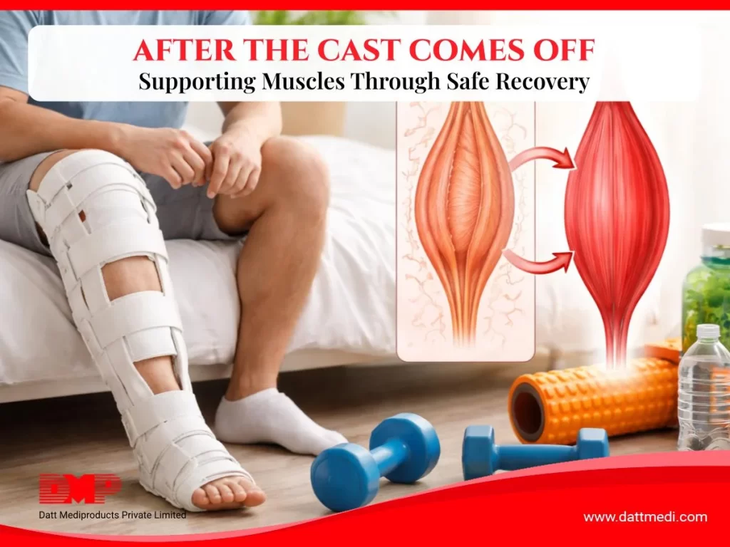
Coming out of a cast, brace, or splint is rarely as straightforward as it looks. The injury may have healed, but the surrounding muscles, joints, and soft tissue have spent weeks in a fixed position with limited activity. What follows is not a return to normal — it is the beginning of a second phase of recovery that requires just as much attention as the first.
This guide covers what actually happens to muscles and joints during immobilization, how to rebuild strength and mobility safely, and which specific orthopedic supports help — matched by body part and stage of recovery.
What immobilization Does to the Body
Muscle reconditioning
Muscles maintain their strength through regular contraction. When a limb is immobilized, the muscles around it stop receiving the mechanical signals they need to stay strong. The result is a measurable loss of muscle mass and endurance — a process called disuse atrophy. Studies show that muscle strength can decline by as much as 3–5% per day in the early stages of immobilization. Even a three-week cast can produce noticeable weakness in the surrounding musculature.
Joint Stiffness and Reduced Range of Motion
Joints rely on movement to stay healthy. Synovial fluid — the lubricant inside joints — circulates through movement. When a joint is held still for weeks, the surrounding capsule and ligaments begin to shorten and stiffen, and the range of motion decreases. Regaining that range requires deliberate, progressive movement — not just returning to normal activity.
Circulation and Swelling
immobilized limbs often develop swelling because the muscle pump that normally drives venous return is inactive. Fluid accumulates in the soft tissue, particularly around the ankle and foot in lower limb immobilization. This swelling can itself delay rehabilitation by limiting movement and causing discomfort.
The Four Phases of Returning to Movement
Phase 1: Protected Range of Motion
Immediately after immobilization ends, the goal is gentle, pain-free movement — not strength. Ankle circles, wrist rotations, finger flexion, and similar small movements reintroduce the joint to its range without stressing the recovering tissue. Movement should stay within a comfortable range and never push into pain.
Phase 2: Circulation and Swelling Control
Before strength can return, swelling needs to reduce. Elevation, gentle movement, and compression where appropriate help manage fluid accumulation. This phase often runs parallel to Phase 1.
Phase 3: Strength Rebuilding
Once range of motion is partly restored and swelling is controlled, progressive resistance exercises can begin. Resistance bands are useful here because they allow graded loading — starting light and increasing gradually without placing sudden stress on healing tissue. Physiotherapy guidance is strongly recommended for this phase.
Phase 4: Functional Reintegration
The final phase returns the person to normal activity — walking, lifting, working, exercising — progressively. Supportive bracing may continue during this phase to provide stability while the muscles are still rebuilding.
The body rebuilds muscle tissue and bone using raw materials from diet. The following nutrients are most directly relevant during post-immobilization recovery:
Protein — essential for muscle repair and rebuilding. Sources include eggs, legumes, dairy, fish, and lean meat. Aim for consistent intake across meals rather than one large serving.
Calcium — supports bone density and structural recovery, particularly relevant after fractures. Dairy products, fortified plant milks, and leafy greens are good sources.
Vitamin D — required for calcium absorption. Many people in India are deficient, particularly those with limited sun exposure. Supplementation is often advisable — check with a doctor.
Collagen precursors (Vitamin C, zinc) — support soft tissue and tendon repair. Found in citrus fruits, berries, nuts, and seeds.
Hydration — adequate water intake supports muscle function, joint lubrication, and circulation during rehabilitation.
Which orthopedic Support to Use — by Body Part and Stage
Supportive braces and sleeves serve two distinct roles during post-immobilization recovery: they protect the recovering joint from excessive or sudden movement, and they provide proprioceptive feedback — helping the nervous system re-learn where the joint is in space. The right product depends on which body part was immobilized and what stage of recovery the person is in.
Supports spinal alignment and shoulder positioning as back and core muscles regain strength
Safe Guidelines for Early Movement
Always Get Clearance First
No exercise or movement programmed should begin without confirmation from the treating clinician or physiotherapist that it is safe to do so. The timeline varies significantly depending on the type of injury, the location, and how healing has progressed.
Start Smaller Than You Think You Need To
The instinct after weeks of immobilization is to move more to make up for lost time. This usually backfires. Muscles and tendons that have been inactive are more vulnerable to strain, and joint tissue is still adapting. Small, controlled movements performed consistently will restore strength faster than pushing into discomfort.
Pain Is a Signal, Not an Obstacle
Discomfort from stiffness is expected and normal. Sharp or worsening pain during exercise is not. If a movement causes significant pain, stop, and raise it with a healthcare professional before continuing.
Use Supports Correctly
A brace or support is most effective when sized and positioned correctly. An ill-fitting support — too loose or too tight — can create pressure points, restrict circulation, or provide false confidence that leads to overexertion. Follow sizing guidance for each product, and if in doubt, seek fitting advice.
When to See a Physiotherapist
Physiotherapy is not just for serious injuries. A physiotherapist can assess the specific degree of muscle weakness and joint restriction after immobilization, design a graduated programmed matched to the person’s actual capacity, and identify any complications — such as tendon tightness, abnormal movement patterns, or persistent swelling — that need addressing before progressing.
The end of immobilization marks the beginning of rebuilding — not the end of recovery. Muscle reconditioning, joint stiffness, and residual swelling are normal consequences of keeping a limb still, and all of them respond well to progressive, consistent rehabilitation.
The right orthopedic support — matched to the body part, the stage of recovery, and the level of activity — makes that process safer and more manageable. Browse the full Datt Mediproducts orthopedic range to find the support that fits your recovery.