Search by Blog
Read by Category

Your healing journey continues beyond this blog

Find the right products for better recovery at online store
Cart Image
The Future of IV Cannulation Velfix® IV Kit

Vascular access in surgical settings is virtually indispensable. The intravenous administration of fluids, medications, blood products and parenteral nutrition, whether peripherally or centrally, is a routine practice in healthcare setups.

The same has been documented in several medical journals such as The American Journal of Epidemiology and the Annals of Pharmacotherapy. According to which, up to 80% of hospitalised patients in recent medical practice receive intravenous therapy while they are admitted.

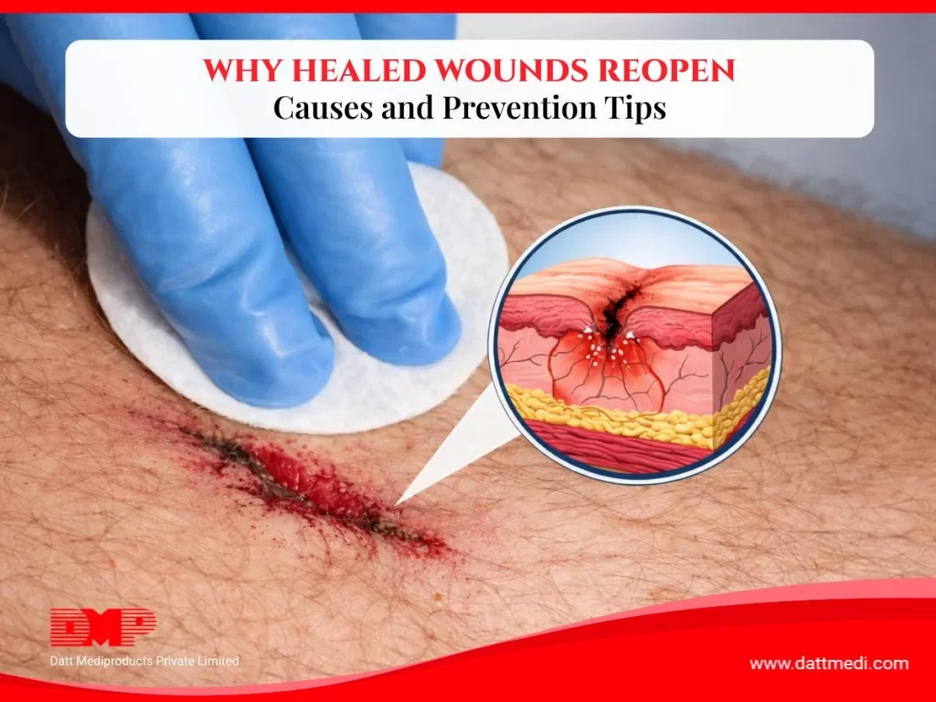
Peripheral intravenous cannulation (PIVC) is the most widely used method for intravenous therapy. Although insertion of cannula and subsequent intravenous therapy are generally well tolerated, complications may arise that may lead to a prolonged hospitalisation. These complications may include Catheter-related bloodstream infections (CR-BSI), Bruising & vein irritation or Blockage. 

Fact:
Epidemiological studies from Europe and US region indicate the incidence of CR-BSI as a percentage of catheters inserted, is between 3% and 7%. (Infections caused by intravascular devices used for infusion therapy: pathogenesis, prevention and management. In: Bison AL, Waldvogel FA, eds. Infections associated with medical devices. Washington DC: ASM Press, 1994:155–205.)

It is vital to understand that any procedure that punctures the skin comes with a risk of associated infections. Since IV sits directly in your bloodstream, a regular observation of the IV site and strict procedures to prevent infection must be practiced by the nursing staff. These will include maintaining good aseptic techniques to minimise the risk of local and systemic infections. 

The CDC Guidelines:

The Centers for Disease Control and Prevention (CDC) have issued certain Guidelines For The Prevention Of Intravascular Catheter-Related Infections. These have been developed for healthcare personnel who insert intravascular catheters and for persons responsible for surveillance and control of infections in hospital, outpatient, and home healthcare settings.

These guidelines include Selection of Catheters; Hand Hygiene & Aseptic Techniques; Maximal Sterile Barrier Precautions; Skin Preparation; Catheter Site Dressing Regimens; Cleansing; Catheter Securement Devices etc.

  • It is recommended to prepare a clean skin with an antiseptic (70% alcohol, tincture of iodine, an iodophor or chlorhexidine gluconate) before peripheral venous catheter insertion.
  • The use of either a sterile gauze or sterile, transparent, semipermeable dressing to cover the catheter site are recommended.
  • It is also suggested to monitor the catheter sites visually when the dressings are changed or by palpation through an intact dressing on a systematically, depending on the clinical situation of each patient. 

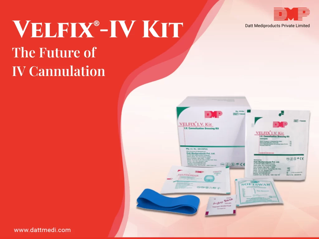
We have introduced a high utility, highly effective IV Cannulization dressing kit specially designed to take care of insertion & dressing change of peripheral vascular devices. 

VELFIX®-IV KIT

A sterile IV Dressing Kit for cannula fixation, including 4 contents in accordance with CDC guidelines.

  • Tourniquet: The kit comes with a latex free tourniquet which helps to identify the veins. Since it is a non-reusable tourniquet, there are a reduced chances of cross- contamination due to carrier property of Velcro tourniquets and thereby preventing skin allergy.
  • Antiseptic Swab: A 70% IPA Swab/chlorhexidine gluconate CHG swab is included in the kit which is considered ideal for skin preparation before peripheral venous catheter insertion. This falls under Category 1B as per Healthcare Infection Control Practices Advisory Committee (HICPAC) implying strong recommendation for implementation, supported by some experimental, clinical, or epidemiologic studies, a strong theoretical rationale; or an accepted practice supported by limited evidence.
  • Velfix® -Edge IV Dressing (7cm x 9cm): Transparent IV dressing with Window Frame delivery design to allow continuous observation of the IV site while providing better seal around the catheter with stabilization non-woven boarders. The notched design ensures effective stabilization of the catheter. The transparent film of the dressing serves as a waterproof sterile barrier to external contaminants. The film is breathable in nature providing high MVTR with an effective wear time of up to 7 days. The dressing comes with a hypoallergenic, latex-free adhesive that is gentle to the skin yet holds catheter in place. It offers single hand application & gentle removal.

The Sterile Gauze Swab of the Velfix® -I.V. Kit is available to control the back flash of blood.

We Datt Mediproducts understand that catheter stabilization is imperative to decrease the risk for phlebitis, catheter migration and dislodgement, and potentially in preventing CR-BSIs. Velfix-IV Kit is an effective and high utility securement device which avoids disruption around the catheter entry site and may reduce the degree of bacterial colonization also. 

Visit www.dattmedi.com for more infomation.
Share:
Recent Blogs
Reopened wounds due to tension and improper care

A wound that looks closed on the surface is not the same as wounds that has finished healing. The skin beneath may still be fragile, thin, and far less resilient than the tissue surrounding it. This is the stage where wounds most commonly reopen — not because something went wrong, but because the final phase of healing was not properly supported.

Understanding what makes newly closed skin vulnerable, and what protective measures actually work, can make the difference between a wound that heals completely and one that breaks down again.

Why ‘Closed Wounds’ Does Not Mean ‘Healed’

Wound healing happens in four overlapping stages: haemostasis, inflammation, proliferation, and remodelling. When the wound appears to close, the body is typically entering the remodelling phase — where collagen fibres reorganise and the new tissue gradually strengthens.

This phase can last several weeks to several months. During this time, the repaired skin has significantly lower tensile strength than normal skin — often as little as 50–80% of the original tissue strength, even after full remodelling. In the early weeks after closure, that figure is even lower.

The practical implication: the skin looks fine but cannot tolerate the same level of stress, friction, or pressure that healthy skin can. This is why reopening happens — not from neglect, but from the invisible vulnerability of newly repaired tissue.

The Most Common Reasons Healed Wounds Reopen

1. Mechanical Stress on Fragile Tissue

Areas that move repeatedly — knees, elbows, ankles, knuckles — place constant tension on healing tissue. Even normal daily movement can stretch new collagen fibres beyond what they can tolerate, causing the wound edges to split. This is particularly common in sutured wounds where the closure relies on the tissue holding together under movement.

2. Friction from Clothing or Surfaces

Repeated low-level friction from fabric, footwear, or contact with surfaces can erode the surface of newly healed skin. Unlike healthy skin, which can handle this without damage, fragile remodelling tissue has little resistance. Wounds on the feet, lower legs, or any area in contact with clothing are especially at risk.

3. Removing Dressings Too Early

Once a wound closes, it is easy to assume that dressings are no longer needed. In practice, premature removal of protective cover exposes the area to exactly the mechanical stress and friction described above. The wound may look healed, but the underlying tissue has not yet reached the strength to handle unprotected exposure.

Overcoming Delayed Wounds Healing

Related reading: Overcoming Delayed Healing: Common Causes and Solutions

4. Moisture Imbalance

Both extremes cause problems. Excess moisture — from wound fluid, sweat, or frequent washing without adequate drying — softens the surrounding skin (maceration), making it far easier to break down under light pressure. Very dry skin, on the other hand, becomes rigid and prone to cracking, particularly over joints. Maintaining a balanced moisture environment under and around the dressing is a key part of protecting fragile tissue.

5. Pressure on Immobile Areas

For individuals with reduced mobility, sustained pressure on a single area — particularly bony prominences like heels, sacrum, or hips — can cause tissue breakdown even in areas that appear to have healed. Regular repositioning and pressure redistribution are essential in these cases.

How Film Dressings Protect Wounds During the Remodelling Phase

Post-operative film dressings are one of the most effective tools for protecting healed and near-healed wounds during the remodelling phase. Unlike gauze-based dressings, which are primarily designed for wounds that are still actively healing and producing exudate, film dressings are specifically suited to closed wounds that need surface protection rather than absorption.

The key properties that make film dressings appropriate for this stage wounds are:

  • Physical barrier — blocks friction, bacteria, dirt, and external mechanical stress
  • Breathability — allows moisture vapour to escape, preventing maceration without drying the tissue
  • Transparency — the wound can be visually monitored without removing the dressing
  • Flexibility — conforms to joints and body contours, maintaining protection during movement
  • Waterproof — the wound stays protected during bathing and daily activity

Datt Mediproducts offers three film and post-operative dressings suited to different stages and wound types in the remodelling phase.

Velfix® T-Film — Transparent Film Dressing

A thin, fully transparent PU film dressing designed for closed wounds that no longer produce exudate. The transparent film allows visual inspection of the wound at any time without disturbing it. Velfix® T-Film is waterproof, breathable, and flexible enough to move with the skin across joints and mobile areas. Available in sizes from 5×7.5 cm to 10×35 cm.

Best for: Sutured wounds, minor cuts and abrasions, and wounds in mobile areas such as knees or elbows that need protection without bulk.

Velfix®-Easy+Pad — Film Dressing with Non-Adherent Pad

Combines a transparent film border with a central non-adherent absorbent pad. This makes Velfix®-Easy+Pad suitable for wounds in the later stages of healing that may still produce minimal fluid, or where the wound surface needs a non-stick contact layer to prevent trauma on removal.

Best for: Post-operative wounds in early recovery that are nearly closed but may have light residual drainage.

Velfix®-T+Pad — Film Dressing with Higher Absorbency

Similar in structure to Easy+Pad but with a higher-absorbency central pad. This is the appropriate choice where some exudate management is still needed alongside the protective film border.

Best for: Wounds transitioning from active healing to the remodelling phase, where light-to-moderate fluid output continues.

Choosing the Right Film Dressing for the Wounds Stage

SituationVelfix® T-FilmEasy+PadT+Pad
Wound fully closed, no fluid✓ IdealSuitableUnnecessary
Minimal residual drainageNot sufficient✓ IdealSuitable
Light-moderate exudateNot suitableBorderline✓ Ideal
Joint or mobile area✓ Ideal✓ Suitable✓ Suitable
Needs visual monitoring✓ IdealPartialPartial

Practical Care Tips for the Remodelling Phase

1. Keep the Area Protected Until the Skin Is Fully Strong

Resist the instinct to leave the area uncovered once the wound looks closed. A film dressing provides protection with minimal bulk and does not interfere with daily activity or bathing.

2. Monitor Without Disturbing

The transparency of film dressings means you can check for redness, fluid accumulation, or early signs of breakdown without peeling back the dressing. Only remove when the dressing edges lift, the dressing is full, or at the interval recommended by your healthcare provider.

3. Watch for Early Warning Signs

Address these promptly rather than waiting to see if they resolve on their own:

  • Redness or warmth spreading from the wound edges
  • Skin that looks shiny, wet, or softened around the dressing
  • Small cracks appearing at the wound edges
  • Increased tenderness or a pulling sensation when moving
  • Any reopening of the wound edges
Infection Control in Wound Care

Related reading: Healing on the Go: Orthopedic and Wound Care Solutions for Physical Activity

Protect the Final Stage as Carefully as the First

Most of the attention in wound care goes to the early stages — cleaning, closing, and dressing the wound. The remodelling phase gets far less focus, but it is where a significant number of wound breakdowns occur. Newly closed skin is fragile, and the stress of normal daily life is often enough to cause reopening without adequate protection.

Film dressings provide a practical, low-profile solution for this stage. Transparent, waterproof, flexible, and gentle on removal — they allow recovery to continue without restricting movement or requiring complex care routines.

Browse the Velfix® T-Film and post-operative dressing range at Datt Mediproducts to find the right option for your stage of recovery.

CHOOSING THE RIGHT DRESSINGS

Chronic wounds are wounds that fail to heal within the expected time frame, typically lasting more than four weeks. They can arise due to underlying conditions such as diabetes, prolonged pressure, or venous insufficiency. Common types include diabetic ulcers, bed sores, and venous leg ulcers.

Managing chronic wounds presents significant challenges, including a high risk of infection, slow healing, and patient discomfort. The right dressing plays a crucial role in creating an optimal healing environment, reducing complications, and improving patient outcomes.

Types of Chronic Wounds and Their Dressing Needs

Diabetic Ulcers

  • A Diabetic foot ulcer is a debilitating complication of diabetes mellitus. It refers  to an open sore or wound, commonly developing on the feet of people with diabetes.
  • Require moisture balance, infection control, and non-adhesive properties to protect fragile skin.

Bed Sores

  • Bed sores are ulcers which usually develop gradually on the skin and the tissue underneath. Occurring mostly in bed ridden patients, bed sores arise on the skin covering bony areas of the body such as hips, back, tailbone, etc.
  • Demand dressings that redistribute pressure and effectively manage exudate to prevent further skin breakdown.

Venous Leg Ulcers

  • Venous leg ulcers are chronic wounds affecting mostly older adults. They primarily result from prolonged venous insufficiency, which leads to tissue breakdown in lower extremities.
  • Benefit from a combination of compression therapy and absorbent dressings to address venous insufficiency and excessive fluid.

Infected Chronic Wounds

  • Chronic infected wounds are those wounds that do not even start to heal after 4 to 12 weeks despite treatment. They develop due to poor blood circulation, weak immune system or co-morbid conditions such as diabetes.
  • Require antimicrobial dressings to manage bacterial load and prevent further complications.

Key Dressing Options:

VEL NeXT™

  • Designed specifically for chronic wounds, including diabetic ulcers, burns, necrotizing fasciitis, and venous ulcers.
  • Promotes faster wound healing while maintaining an optimal moisture balance.

Velvert®

  • A secure antimicrobial dressing with excellent moisture management.
  • Ideal for wounds at high risk of infection, including diabetic ulcers and chronic wounds.

Silvel™

  • Features nano-crystalline silver for strong antimicrobial action.
  • Features silver for strong antimicrobial action.
  • Suitable for infected or high-risk wounds, particularly bed sores.

Velsap™

  • Designed for moderate to heavily exuding wounds.
  • Provides effective fluid management, reducing the need for frequent dressing changes.

How to Choose the Right Dressing?

Selecting the right dressing requires a comprehensive assessment of several factors:

  • Wound Type: Identify whether the wound is a diabetic ulcer, pressure sore, or venous ulcer.
  • Exudate Levels: Choose absorbent dressings for heavily exuding wounds and moisture-retaining options for dry wounds.
  • Infection Status: Use antimicrobial dressings for infected or high-risk wounds.
  • Patient-Specific Considerations: Factors such as mobility, skin sensitivity, and underlying health conditions must be considered.
Healthcare professionals play a crucial role in guiding dressing selection based on individual wound characteristics and patient needs.

Additional Tips for Chronic Wound Care Management

  • Proper Cleaning & Debridement: Keeping the wound clean and removing dead tissue enhances healing.
  • Regular Monitoring & Dressing Changes: Assessing the wound regularly and changing dressings at the right intervals prevent complications and ensure steady healing.

Chronic wounds require tailored solutions to support healing and prevent complications. Choosing the right dressing—ensures optimal wound care and faster recovery.

Explore our advanced dressing solutions today and find the best option for your wound care needs!

Logo

Datt Mediproducts Private Limited

Loading